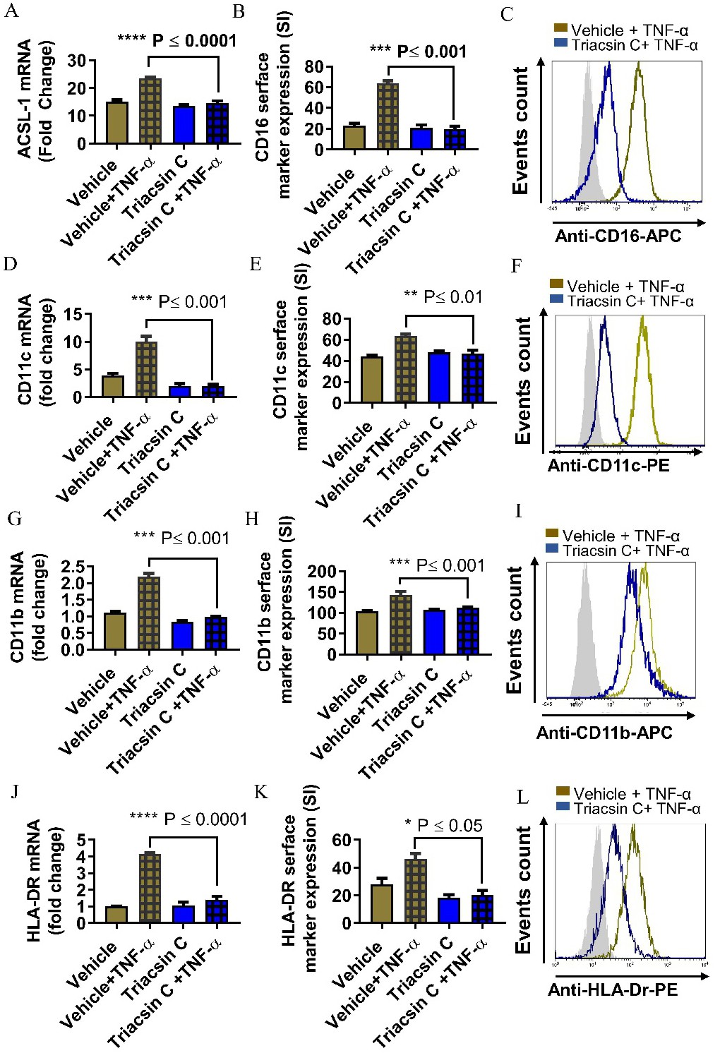
Fig. 1. ACSL1 inhibition blocks TNF-α mediated pro-inflammatory change in monocytes. Monocytic cells were pretreated with ACSL1 inhibitor (Triacsin C: (1uM) or vehicle for 1 hour and then incubated with TNF-α for 24 hours. (A, D, G, J) ACSL, CD11c, CD11b and HLA-DR mRNA were determined by real time RT-PCR. Triacsin C inhibted Cells were stained with antibody against CD16, CD11c, CD11b, or HLA-DR, along with matched control antibodies and assessed by flow cytometry. (B, E, H, K) Flow cytometry data are presented as a bar graph of mean staining intensity (SI) and (C, F, I, L) representative histogram. Bar graphs depict mean values ± SEM of staining intensity (SI). P<0.05 was considered as statistically significant (* P<0.01; **P< 0.001, ***P< 0.0001). The data in all figures are representative of three independent experiments.